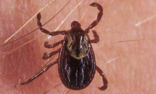
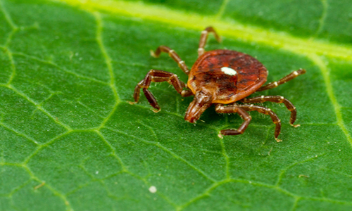
Our previous post provided information about diseases and pathogens spread by Blacklegged (Deer) Tick bites. In this post, we are focusing on those diseases which are commonly spread by American Dog ticks, largest of all ticks found in the Northeast. The adult female American Dog ticks have a white horseshoe characteristic while the males appear to have lightning bolts of white across their back sides. The Lone Star ticks are slightly smaller with the adult females featuring a distinctive white dot on their dorsal shield. Both of these ticks are capable of transmitting bacterial and viral infections, specifically, we’ll be discussing Ehrlichiosis, Rocky Mountain Spotted Fever and Tularemia.
If you should experience or know of anyone experiencing these types of symptoms, quickly follow up with a medical professional.
Ehrlichiosis (Ehrlichia chaffeensis & Ehrlichia ewingii)
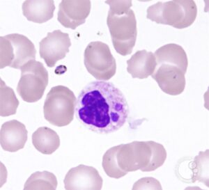
American Dog ticks and Lone Star ticks are known to spread the two different species of Ehrlichiosis (Ehrlichia chaffeensis and Ehrlichia ewingii). Transmission time from bite to infection is between 18-24 hours. Both species invade the white blood cells, causing Human Monocytic Ehrlichiosis with symptoms appearing 1-2 weeks after exposure.
Ehrlichiosis symptoms can include chills, sweats, headache, fatigue, nausea, diarrhea, vomiting, fever, joint and muscle pain, disorientation, rash and eye infection. Immunocompromised individuals may also experience trouble breathing and kidney dysfunction. Even after a few weeks of treatment, headache and fatigue symptoms may continue to be present.
Polymerase Chain Reaction (PCR) of the blood within the first week of infection can detect Ehrlichia DNA while peripheral blood smears may also show Ehrlichia in white blood cells confirming diagnosis. Antibodies can also be detected using a two-part Indirect Immunofluorescence Assays (IFA) test. Finally, low platelet and white blood cell counts or elevated liver enzymes may suggest Ehrlichiosis infection.
Rocky Mountain Spotted Fever (Rickettsia amblyommii, Rickettsia rickettsia & Rickettsia parkeri)
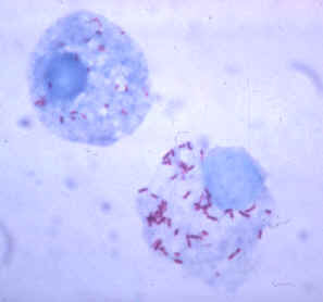
The American Dog ticks and Lone Star ticks may transmit a bacterium causing Rocky Mountain Spotted Fever which is known as a Rickettsial bacterial. It infects the cells that line blood vessels causing vascular inflammation known as Rickettsial vasculitis. Between 6-10 hours this bacterium can pass from bite to infection and symptoms typically develop between 2-12 days later. A spotted rash varying in presentation will occur in most patients.
Rocky Mountain Spotted Fever symptoms can include headache, fever, nausea and vomiting, and can be followed by a quick progression into more severe and life-threatening symptoms. Blood vessel damage may lead to amputation if left untreated, and mental and physical disability can occur in severe cases.
Immunostaining of skin biopsies can confirm the presence of Rickettsia amblyommii & Rickettsia spp and antibodies can be detected through a two-part Indirect Immunofluorescence Assays (IFA) test. The first sample is collected within 7 days of infection and will be compared to the second sample collected 2-4 weeks after infection. If the second sample shows the number of antibodies present has increased since the first sample was tested, the results are positive for Rickettsia. False positives are common for the first part of the test. Finally, blood test results of low platelet count, low sodium levels or elevated liver enzyme levels can assist with diagnosis.
Tularemia (Francisella tularensis)
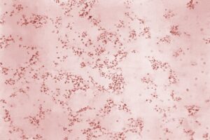
Finally, American Dog tick and Lone Star Tick bites can spread a bacterial infection which, 3-5 days after the initial bite, can cause Tularemia.
Tularemia symptoms can range from mild to life-threatening and commonly include high fever. Glandular infections occur when the lymph nodes closest to the tick bite are swollen and a high fever is present. Ulcerglandular infections occur when the lymph nodes closest to the tick bite are swollen, a high fever is present, and there is a skin ulcer at the site of tick attachment.
For diagnosis, skin biopsy or secretion can be visualized under a microscope using a gram stain or fluorescently labelled antibodies to identify the presence of Francisella tularensis.
Again, if you experience or know of someone experiencing any of these symptoms, please follow up and consult with a medical professional.
Resources:
